
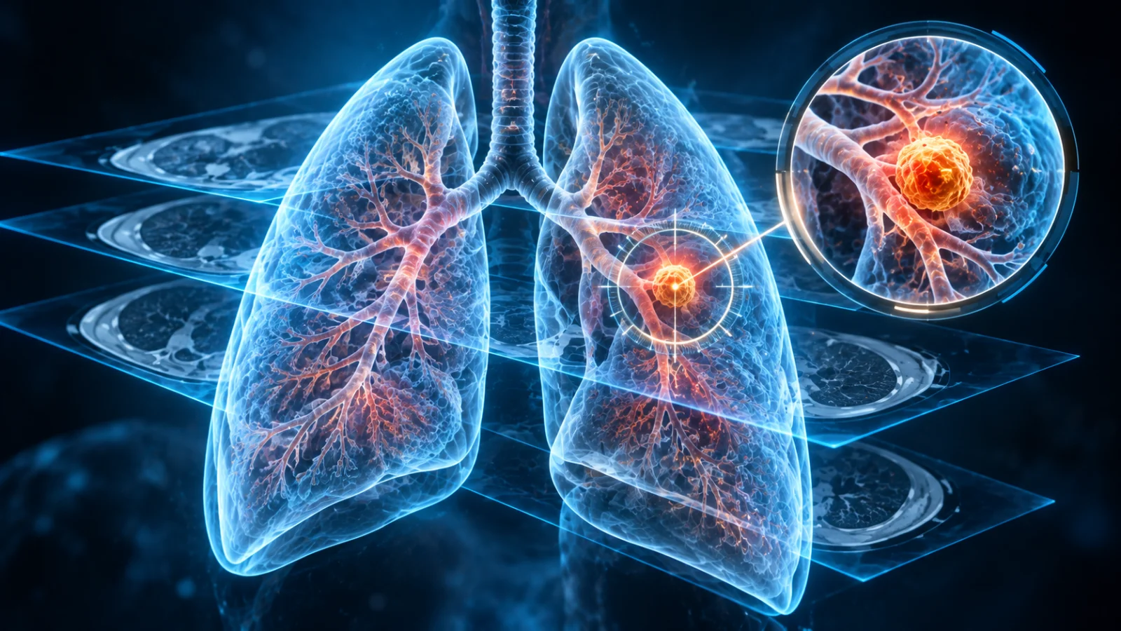
You went for a CT scan for something else entirely — a nagging cough, a health check-up, a kidney stone, a minor accident. Then one line in the report jumps out: “A small nodule is noted in the right lung.” Suddenly that’s all you can think about.
Lakshmi (name changed), a 52-year-old teacher from Miyapur, had a CT scan after a bout of pneumonia. The report mentioned a 7 mm nodule. She spent three sleepless nights searching online, convinced she had lung cancer.
When she brought her scans in, we compared them with a chest CT she had taken during COVID four years earlier. The nodule was already there — exactly the same size. A solid nodule that hasn’t grown in over two years is almost always benign. Hers was most likely an old, healed infection scar. She walked out relieved, with a simple plan.
Lakshmi’s fear is completely understandable. But here is the most important thing to know about a lung nodule: the vast majority are not cancer. What matters is having a clear, sensible plan — not panic, and not neglect.
Key takeaways
- A lung nodule is a round spot up to 3 cm across; anything larger is called a “mass”.
- Nodules are very common on CT scans, and most are benign — especially in India, where old TB and fungal infections leave harmless scars.
- Small solid nodules under 6 mm carry a very low cancer risk (well under 1% in most people).
- Size, shape, growth over time and your personal risk factors decide what happens next.
- Your old scans are gold — always bring previous X-rays and CTs to your appointment.
What exactly is a lung nodule?
A lung nodule (also called a pulmonary nodule or “coin lesion”) is a small, roughly round area that looks denser than the surrounding lung on an X-ray or CT scan. Nodules usually cause no symptoms at all — which is why they are almost always discovered by accident, on a scan done for another reason.
Modern CT scanners are so sensitive that they pick up spots just 2–3 mm across. That is a good thing for early detection, but it also means many people are now told about tiny nodules that would never have caused them any harm.
What causes lung nodules?
5 clues doctors use to judge a nodule
1Size
Generally, the smaller the nodule, the lower the risk. Tiny nodules under 6 mm are very rarely cancerous.
2Growth over time
This is often the single most reassuring clue. A solid nodule that stays the same size for two years is considered benign. That’s why comparing with old scans — like Lakshmi’s — can settle the question instantly.
3Shape and edges
Smooth, round edges and certain patterns of calcium (like a bull’s-eye or “popcorn” pattern) suggest a harmless nodule. Irregular, spiky (“spiculated”) edges raise more concern.
4Type: solid or “ground-glass”
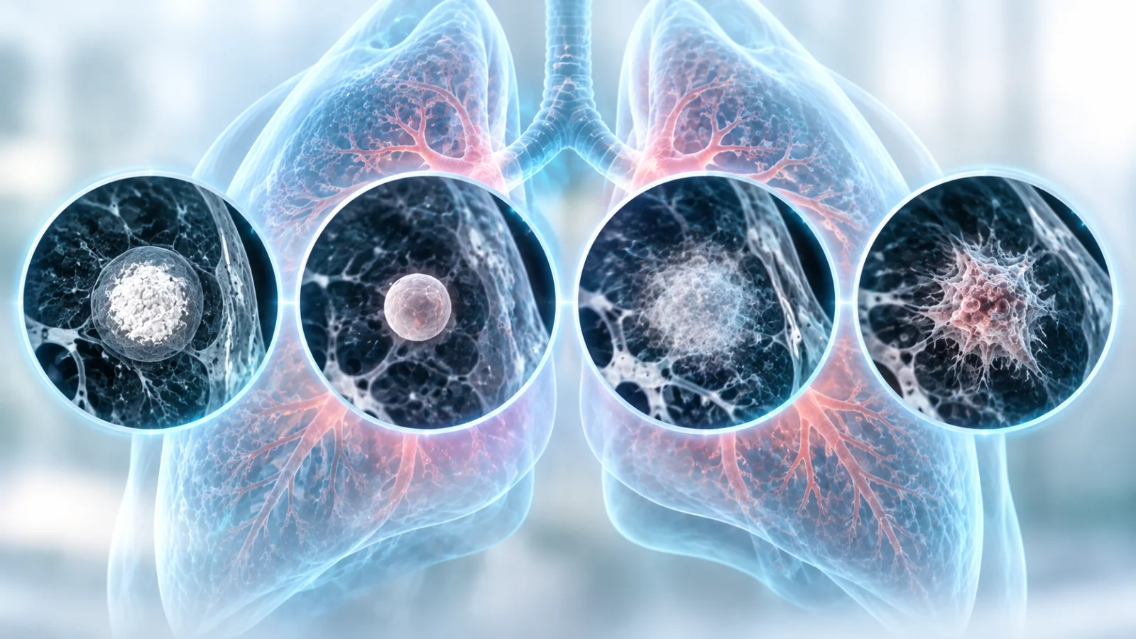
Some nodules look hazy, like frosted glass, rather than solid white. These ground-glass or part-solid nodules often fade away (if caused by inflammation) but, when they persist, they can be very slow-growing early cancers — so they are followed for longer, sometimes up to five years.
5You — your personal risk
Age over 50, current or past smoking, a family history of lung cancer, exposure to asbestos, silica or heavy smoke (including biomass cooking fuel), and previous cancers all raise the level of caution.
Nodule size and what usually happens next
Radiologists and pulmonologists worldwide use guidelines (such as those from the Fleischner Society) to decide follow-up for nodules found by chance. The table below is a simplified overview for a single solid nodule in an adult — your doctor will tailor the plan to your scan and history.
| Nodule size | Typical risk | Usual next step |
|---|---|---|
| Under 6 mm | Very low | Often no routine follow-up in low-risk people; an optional repeat CT at 12 months for higher-risk people. |
| 6–8 mm | Low | Repeat CT in about 6–12 months, then again later depending on findings and risk. |
| Over 8 mm | Low to moderate | Consider a repeat CT at around 3 months, a PET-CT scan, or a biopsy. |
| Over 3 cm (a “mass”) | Higher | Prompt evaluation, usually including biopsy. |
Why we don’t biopsy everything: Testing every tiny nodule would expose thousands of people to procedures, radiation and anxiety for spots that are harmless. Careful “watchful waiting” with low-dose CT is a deliberate, evidence-based strategy — not neglect.
When is a biopsy needed — and how is it done?
If a nodule is large, growing, or looks suspicious, getting a tissue sample is the only way to know for sure. Today, many biopsies are done without open surgery:
- PET-CT scan — shows how “active” a nodule is. In India, one caution: TB and other infections can also light up on PET, so a bright result isn’t automatically cancer.
- CT-guided needle biopsy — a thin needle is guided through the chest wall into the nodule.
- Bronchoscopy with radial EBUS — a flexible camera is passed through the mouth into the airways, and a tiny ultrasound probe locates the nodule from inside. No cuts on the skin. Learn about linear and radial EBUS.
- EBUS-TBNA — samples enlarged lymph nodes in the chest, which helps both diagnose and stage lung cancer in one sitting.
- Cryobiopsy — uses extreme cold to take larger, better-quality samples. See cryo lung biopsy.
These minimally invasive procedures are typically done under sedation, often as a day-care procedure. If a nodule does turn out to be cancer, finding it at this small stage offers the best chance of complete cure. Read more about our approach to lung cancer management.
Myths that cause unnecessary fear
Don’t wait — see a pulmonologist soon if
- The nodule is larger than 8 mm, or has grown since a previous scan
- The report mentions “spiculated”, “irregular” or “suspicious” features
- You have symptoms such as coughing up blood, persistent cough, weight loss or chest pain
- You are a current or former smoker over 50
Questions to ask your doctor
- How big is the nodule, and is it solid or ground-glass?
- Can you compare it with my older scans?
- Based on my risk factors, how likely is it to be benign?
- When exactly should my next scan be — and should it be a low-dose CT?
- What findings would make us move to a biopsy?
Write the follow-up date in your calendar and on your phone. A missed follow-up scan is one of the most common — and most avoidable — problems with lung nodules.
Frequently asked questions
What percentage of lung nodules are cancerous?
Can a lung nodule go away on its own?
Should I be worried about a 4 mm lung nodule?
Can tuberculosis cause lung nodules?
Do lung nodules cause symptoms?
Worried about a nodule on your scan?
Bring your CT report and any older scans. Dr. Subhakar Nadella will review them with you, explain exactly what your nodule means, and create a clear follow-up or biopsy plan — using minimally invasive techniques like EBUS and cryobiopsy when needed.

Dr. Subhakar Nadella
MD (Pulmonary Medicine) · Consultant Clinical & Interventional Pulmonologist, KIMS Hospital, Kondapur, Hyderabad
With over 10 years in pulmonary medicine — including more than 4 years in critical care and advanced interventions — Dr. Nadella treats the full spectrum of lung and breathing problems, from chronic cough, asthma and COPD to sleep apnea, interstitial lung disease, lung nodules and lung cancer.
- Flexible & rigid bronchoscopy
- Linear & radial EBUS
- Cryo lung biopsy
- Medical thoracoscopy
- Foreign body removal
- Sleep disorders
- Critical care
This article is for general education and does not replace a personal medical consultation. Follow-up recommendations vary with individual risk; please consult a qualified doctor about your scan. Patient names and details have been changed to protect privacy. Reference: MacMahon H, et al. Guidelines for Management of Incidental Pulmonary Nodules Detected on CT Images: From the Fleischner Society 2017. Radiology. 2017.